Αρχειοθήκη ιστολογίου

Αλέξανδρος Γ. Σφακιανάκης
ΩτοΡινοΛαρυγγολόγος
Αναπαύσεως 5
Άγιος Νικόλαος Κρήτη 72100
2841026182
6032607174

Τετάρτη 14 Οκτωβρίου 2020

Flow-Induced Transcriptomic Remodeling of Endothelial Cells Derived From Human Induced Pluripotent Stem Cells

Flow-Induced Transcriptomic Remodeling of Endothelial Cells Derived From Human Induced Pluripotent Stem Cells:

591450_Thumb_400.jpg


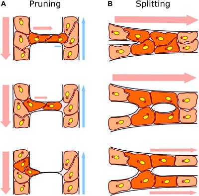
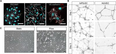
The vascular system is essential for the development and function of all organs and tissues in our body. The molecular signature and phenotype of endothelial cells (EC) are greatly affected by blood flow-induced shear stress, which is a vital component of vascular development and homeostasis. Recent advances in differentiation of ECs from human induced pluripotent stem cells (hiPSC) have enabled development of in vitro experimental models of the vasculature containing cells from healthy individuals or from patients harboring genetic variants or diseases of interest. Here we have used hiPSC-derived ECs and bulk- and single-cell RNA sequencing to study the effect of flow on the transcriptomic landscape of hiPSC-ECs and their heterogeneity. We demonstrate that hiPS-ECs are plastic and they adapt to flow by expressing known flow-induced genes. Single-cell RNA sequencing showed that flow induced a more homogenous and homeostatically more stable EC population compared to static cultures, as genes related to cell polarization, barrier formation and glucose and fatty acid transport were induced. The hiPS-ECs increased both arterial and venous markers when exposed to flow. Interestingly, while in general there was a greater increase in the venous markers, one cluster with more arterial-like hiPS-ECs was detected. Single-cell RNA sequencing revealed that not all hiPS-ECs are similar even after sorting, but exposing them to flow increases their homogeneity. Since hiPS-ECs resemble immature ECs and demonstrate high plasticity in response to flow, they provide an excellent model to study vascular development.

Two Undervalued Functions of the Golgi Apparatus: Removal of Excess Ca2+ and Biosynthesis of Farnesol-Like Sesquiterpenoids, Possibly as Ca2+-Pump Agonists and Membrane “Fluidizers–Plasticizers”

Two Undervalued Functions of the Golgi Apparatus: Removal of Excess Ca2+ and Biosynthesis of Farnesol-Like Sesquiterpenoids, Possibly as Ca2+-Pump Agonists and Membrane “Fluidizers–Plasticizers”:

542879_Thumb_400.jpg


The extensive literature dealing with the Golgi system emphasizes its role in protein secretion and modification, usually without specifying from which evolutionary ancient cell physiological necessity such secretion originated. Neither does it specify which functional requirements the secreted proteins must meet. From a reinterpretation of some classical and recent data gained mainly, but not exclusively, from (insect) endocrinology, the view emerged that the likely primordial function of the rough endoplasmic reticulum (RER)–Golgi complex in all eukaryotes was not the secretion of any type of protein but the removal of toxic excess Ca2+ from the cytoplasm. Such activity requires the concurrent secretion of large amounts of Ca2+-carrying/transporting proteins acting as a micro-conveyor belt system inside the RER–Golgi. Thus, (fitness increasing) protein secretion is subordinate to Ca2+ removal. Milk with its high content of protein and Ca2+ (60–90 mM vs. 100 nM in unstimulated mammary gland cells) is an extreme example. The sarco(endo)plasmatic reticulum Ca2+-ATPases (SERCAs) and SPCA1a Ca2+/Mn2+ transport ATPases are major players in Ca2+ removal through the Golgi. Both are blocked by the sesquiterpenoid thapsigargin. This strengthens the hypothesis (2014) that endogenous farnesol-like sesquiterpenoids (FLSs) may act as the long sought for but still unidentified agonist(s) for Ca2+-pumps in both the ER and Golgi. A second putative function also emerges. The fusion of both the incoming and outgoing transport vesicles, respectively, at the cis- and trans- side of Golgi stacks, with the membrane system requiring high flexibility and fast self-closing of the involved membranes. These properties may—possibly partially—be controlled by endogenous hydrophobic membrane “fluidizers” for which FLSs are prime candidates. A recent reexamination of unexplained classical data suggests that they are likely synthesized by the Golgi itself. This game-changing hypothesis is endorsed by several arguments and data, some of which date from 1964, that the insect corpus allatum (CA), which is the major production site of farnesol-esters, has active Golgi systems. Thus, in addition to secreting FLS, in particular juvenile hormone(s), it also secretes a protein(s) or peptide(s) with thus far unknown function. This paper suggests answers to various open questions in cell physiology and general endocrinology.

Physical Exercise and the Renin Angiotensin System: Prospects

Physical Exercise and the Renin Angiotensin System: Prospects in the COVID-19:

561403_Thumb_400.jpg


Recent reports have shown that the renin angiotensin system (RAS) plays an important role in the Coronavirus disease 2019 (COVID-19) because the angiotensin converting enzyme 2 is the receptor for the severe acute respiratory syndrome coronavirus 2. In addition, the balance of RAS components can be involved in the pathogenesis and progression of COVID-19, especially in patients with metabolic and cardiovascular diseases. On the other hand, physical exercise is effective to prevent and to counteract the consequences of such diseases and one of the biological mediators of the exercise adaptation is the RAS. This review was designed to highlight the connection between COVID-19 and RAS, and to discuss the role of the RAS as a mediator of the benefits of physical exercise in COVID-19 pandemic.

Functional Characterization of Facilitative Glucose Transporter 4 With a Delay Responding to Plasma Glucose Level in Blunt Snout Bream (Megalobrama amblycephala)

Functional Characterization of Facilitative Glucose Transporter 4 With a Delay Responding to Plasma Glucose Level in Blunt Snout Bream (Megalobrama amblycephala):

582785_Thumb_400.jpg


Facilitative glucose transporter 4 (GLUT4) plays a central role in mediating insulin function to increase glucose uptake in glucose metabolism homeostasis. In this study, the function and localization of GLUT4 in blunt snout bream (Megalobrama amblycephala) were first investigated, and then, the response measured as carbohydrate level, was analyzed. The results showed that the cDNA sequence of GLUT4 in blunt snout bream (MaGLUT4, GenBank accession no: MT447093) was 2868 bp in length, and the corresponding mRNA contained a 5′-UTR region of 513 bp and a 3′-UTR region of 837 bp. MaGLUT4 had an open reading frame of 1518 bp and was encoded by 505 amino acids. Its theoretical isoelectric point and molecular weight was 6.41 and 55.47 kDa, respectively. A comparison of these characteristics with BLASTP results from the NCBI database showed that MaGLUT4 had the highest homology with Cypriniformes fish, with MaGLUT4 and GLUT4 of other Cypriniformes clustered in the phylogenetic tree with other GLUT1–4 amino acid sequences. Compared with the results from the homo_sapiens and mus_musculus data sets, some mutations were observed in the GLUT4 amino acid sequence of these aquatic animals, including an FQQI mutation to FQQL, LL mutation to MM, and TELEY mutation to TELDY. MaGLUT4 was constitutively expressed in the muscle, intestine, and liver, with the highest mRNA level observed in muscle. Furthermore, the predicted tertiary structure and results of immunohistochemical staining showed that MaGLUT4 was a transmembrane protein primarily located in the plasma membrane, where it accounts for 60.9% of the total expressed, according to an analysis of subcellular localization. Blood glucose level peaked within 1 h, and the insulin level peaked at 6 h, while the mRNA and protein levels of GLUT4 showed an upward trend with an increase in feeding time and decreased sharply after 12 h. These results confirmed that MaGLUT4 was mainly distributed in muscles and crosses the cell membrane. The changes in the insulin, mRNA, and protein levels of MaGUT4 lagged far behind changes in blood glucose levels. This delay in insulin level changes and GLUT4 activation might be the important reasons for glucose intolerance of this fish species.

Palmitoyl Protein Thioesterase 1 Is Essential for Myogenic Autophagy of C2C12 Skeletal Myoblast

Palmitoyl Protein Thioesterase 1 Is Essential for Myogenic Autophagy of C2C12 Skeletal Myoblast:

569221_Thumb_400.jpg


Skeletal muscle differentiation is an essential process for the maintenance of muscle development and homeostasis. Reactive oxygen species (ROS) are critical signaling molecules involved in muscle differentiation. Palmitoyl protein thioesterase 1 (PPT1), a lysosomal enzyme, is involved in removing thioester-linked fatty acid groups from modified cysteine residues in proteins. However, the role of PPT1 in muscle differentiation remains to be elucidated. Here, we found that PPT1 plays a critical role in the differentiation of C2C12 skeletal myoblasts. The expression of PPT1 gradually increased in response to mitochondrial ROS (mtROS) during muscle differentiation, which was attenuated by treatment with antioxidants. Moreover, we revealed that PPT1 transactivation occurs through nuclear factor erythroid 2-regulated factor 2 (Nrf2) binding the antioxidant response element (ARE) in its promoter region. Knockdown of PPT1 with specific small interference RNA (siRNA) disrupted lysosomal function by increasing its pH. Subsequently, it caused excessive accumulation of autophagy flux, thereby impairing muscle fiber formation. In conclusion, we suggest that PPT1 is factor a responsible for myogenic autophagy in differentiating C2C12 myoblasts.

Remodeling of the Microvasculature

Remodeling of the Microvasculature: May the Blood Flow Be With You:

586852_Thumb_400.jpg


The vasculature ensures optimal delivery of nutrients and oxygen throughout the body, and to achieve this function it must continually adapt to varying tissue demands. Newly formed vascular plexuses during development are immature and require dynamic remodeling to generate well-patterned functional networks. This is achieved by remodeling of the capillaries preserving those which are functional and eliminating other ones. A balanced and dynamically regulated capillary remodeling will therefore ensure optimal distribution of blood and nutrients to the tissues. This is particularly important in pathological contexts in which deficient or excessive vascular remodeling may worsen tissue perfusion and hamper tissue repair. Blood flow is a major determinant of microvascular reshaping since capillaries are pruned when relatively less perfused and they split when exposed to high flow in order to shape the microvascular network for optimal tissue perfusion and oxygenation. The molecular machinery underlying blood flow sensing by endothelial cells is being deciphered, but much less is known about how this translates into endothelial cell responses as alignment, polarization and directed migration to drive capillary remodeling, particularly in vivo. Part of this knowledge is theoretical from computational models since blood flow hemodynamics are not easily recapitulated by in vitro or ex vivo approaches. Moreover, these events are difficult to visualize in vivo due to their infrequency and briefness. Studies had been limited to postnatal mouse retina and vascular beds in zebrafish but new tools as advanced microscopy and image analysis are strengthening our understanding of capillary remodeling. In this review we introduce the concept of remodeling of the microvasculature and its relevance in physiology and pathology. We summarize the current knowledge on the mechanisms contributing to capillary regression and to capillary splitting highlighting the key role of blood flow to orchestrate these processes. Finally, we comment the potential and possibilities that microfluidics offers to this field. Since capillary remodeling mechanisms are often reactivated in prevalent pathologies as cancer and cardiovascular disease, all this knowledge could be eventually used to improve the functionality of capillary networks in diseased tissues and promote their repair.

Effect of Hyperbilirubinemia on Medial Olivocochlear System in Newborns.

Effect of Hyperbilirubinemia on Medial Olivocochlear System in Newborns.:

Icon for Aves Yayincilik Icon for PubMed Central

Logo of jiaoLink to Publisher's site
. 2020 Aug; 16(2): 295–296.
PMCID: PMC7419097
PMID: 32784171

Effect of Hyperbilirubinemia on Medial Olivocochlear System in Newborns

corresponding authorCorresponding author.
Corresponding Address: Fulya Özer, E-mail: moc.liamtoh@60icevedf
Dear Editor,
I have read with great interest the article titled “Effect of Hyperbilirubinemia on Medial Olivocochlear System in Newborns” in the August 2019 issue of The Journal of International Advanced Otology (15(2): 272–6) by Karabulut et al. [] However, some points need to be explained.
The authors demonstrated subclinical damage in Medial Olivocochlear System (MOC) efferent system of infants with hyperbilirubinemia with normal auditory brain stem response. They showed that MOC reflex activity significantly decreased with hyperbilirubinemia. They also claimed that there was no significant correlation between total serum bilirubin levels and total MOC reflex values of both ears and presented some p values. I could not find the mean values of total serum bilirubin levels and of total MOC reflex. It is not clear which statistical test is used for this insignificant relationship. I assume that they performed a correlation analysis, as they say there is no correlation.
However, correlation analysis is a statistical method used to determine whether there is a linear relationship between two (or more) quantitative variables []. Correlation coefficient is used for this statistic and expressed as “r.” If r < 0.2, it means there is no correlation or weak correlation. In the article by Karabulut et al., it should be the r coefficient, not the p value, which should be presented for statistical significance, and this relationship should be shown with a graph.
In the literature, Jiang et al. [] showed that there was no close correlation between the degree of amplitude reduction in auditory brainstem response (ABR) and the level of total serum bilirubin. However, they found significant correlation between latencies of ABR and total serum bilirubin level in another study []. Amin et al. [] claimed that the best indicator for the toxic effect of hyperbilirubinemia on the hearing system is the level of indirect bilirubin level not total serum bilirubin. In the literature, there is no information about the relationship between MOC efferent system and total bilirubin level or indirect bilirubin level as mentioned by authors.
Therefore, I believe that not being able to find a relationship between MOC reflex activity and total serum bilirubin level is a very important conclusion with high citation probability. This result will be also essential for future studies about the relationship between MOC reflex activity and indirect bilirubin levels. However, this conclusion should be proven with the correct statistics, data, and graphics.

Footnotes

Peer-review: Externally peer-reviewed.
Conflict of Interest: The author has no conflict of interest to declare.
Financial Disclosure: The author declared that this study has received no financial support.

REFERENCES

1. Karabulut B, Sürmeli M, Bozdağ Ş, Deveci İ, Doğan R, Oysu Ç. Effect of Hyperbilirubinemia on Medial Olivocochlear System in Newborns. J Int Adv Otol. 2019;15:272–6. doi: 10.5152/iao.2019.5723. [PMC free article] [PubMed] [CrossRef[]
2. Gogtay NJ, Thatte UM. Principles of Correlation Analysis. J Assoc Phsicians India. 2017;65:78–81. [PubMed[]
3. Jiang ZD, Brosi DM, Wilkinson AR. Changes in BAER wave amplitudes in relation to total serum bilirubin level in term neonates. Eur J Pediatr. 2009;168:1243–50. doi: 10.1007/s00431-008-0919-3. [PubMed] [CrossRef[]
4. Jiang ZD, Chen C, Liu TT, Wilkinson AR. Changes in Brainstem Auditory EvokedResponse Latencies in Term Neonates with Hyperbilirubinemia. Pediatr Neurol. 2007;37:35–41. doi: 10.1016/j.pediatrneurol.2007.03.006. [PubMed] [CrossRef[]
5. Amin SB, Saluja S, Saili A, Orlando M, Wang H, Laroia N, et al. Chronic Auditory Toxicity in Late Preterm and Term Infants with Significant Hyperbilirubinemia. Pediatrics. 2017;40:e20164009. doi: 10.1542/peds.2016-4009. [PMC free article] [PubMed] [CrossRef[]

Articles from The Journal of International Advanced Otology are provided here courtesy of The European Academy of Otology and Neurotology