Αρχειοθήκη ιστολογίου

Αλέξανδρος Γ. Σφακιανάκης
ΩτοΡινοΛαρυγγολόγος
Αναπαύσεως 5
Άγιος Νικόλαος Κρήτη 72100
2841026182
6032607174

Πέμπτη 14 Ιανουαρίου 2021

A Complication of Mycobacterium chelonae Infection Following Orbital Floor Fracture Repair

xloma.fota13 shared this article with you from Inoreader

SocialThumb.00001665.DC.jpeg

Mycobacterium chelonae is a rare, atypical nontuberculous bacterium that has been reported to be an underlying cause of persistent wound infections. Although there are several studies highlighting the role of M chelonae as the putative cause of other postoperative wound infections, to our knowledge there ar e no reports of infection following implant placement for repair of an orbital floor fracture. The authors present a unique case describing the management of a persistent postoperative infection in a young, immunocompetent patient with an orbital floor fracture repaired with a Stryker Medpor Titan implant. The patient was initially treated with broad-spectrum antibiotics with minimal clinical improvement. Following culture-proven M chelonae, a second surgical intervention was undertaken to remove the implant and later, a third intervention for scar revision. The patient has remained free of infection utilizing a long-term tailored 2-drug antibiotic regimen. This case emphasizes the need for recognition of M chelonae as a potential pathogen in certain clinical situations and the difficulty in eradicating M chelonae in the context of infected implantable devices. The comprehensive treatment protocol required to ensure adequate therapy is reviewed. Address correspondence and reprint requests to J. Madison Clark, MD, Department of Otolaryngology-Head and Neck Surgery, Division of Facial Plastic and Reconstructive Surgery, University of North Carolina Hospitals, 170 Manning Dr., Campus Box #7070, Chapel Hill, NC 27599; E-mail: Madison_clark@med.unc.edu Received 9 September, 2020 Accepted 3 December, 2020 This study was approved by the Institutional Review Board at the University of North Carolina Chapel Hill (20-0558). The authors report no conflicts of interest. © 2021 by Mutaz B. Habal, MD.
View on the web

Outcomes in Orbital Floor Trauma: A Comparison of Isolated and Zygomaticomaxillary-Associated Fractures

xloma.fota13 shared this article with you from Inoreader

SocialThumb.00001665.DC.jpeg

Orbital floor fractures are common sequalae of trauma to the orbit. These fractures present as an isolated orbital floor (I-OF) fracture or with other midface fractures, typically the zygomaticomaxillary complex. The authors sought to better understand the differences in patient presentation, surgical decis ion-making, and outcomes in I-OF fractures compared with those associated with zygomaticomaxillary complex fractures (Z-OF). A retrospective review of patients with orbital floor fractures was conducted to generate an I-OF fracture group and a Z-OF fracture group. Demographics, preoperative symptoms, surgical choices, and postoperative complications were assessed. Complications were assessed individually and as 2 composite groups consisting of orbital complications and eyelid complications. There were 156 patients that met inclusion criteria with 75 I-OF fractures and 81 Z-OF fractures. The most common mechanism of injury for I-OF fractures was assault (34.7%) and motor vehicle accidents (39.5%) for Z-OF fractures. The I-OF group presented more often with diplopia (P = 0.01) whereas the Z-OF group had more trauma symptoms (P = 0.01), which included subconjunctival hemorrhages, retrobulbar hemorrhages, and relative afferent pupillary defects. I-OF fractures had longer preoper ative observational periods (P 
View on the web

Facial Fractures and Mixed Dentition – What Are the Implications of Dentition Status in Pediatric Facial Fracture Management?

xloma.fota13 shared this article with you from Inoreader

SocialThumb.00001665.DC.jpeg

Introduction: The stage of maturation of the pediatric facial skeleton at the time of injury has a significant impact on both facial fracture patterns and management strategies. For instance, the relative prominence of the pediatric cranium during the early years of life affords protection to the structures of the midface, whereas delayed aeration of the frontal sinuses may predispose younger patients to frontal bone fractures. The dentition status of a pediatric patient may have similar implications in the setting of facial fracture. In this study, the authors examine the effect of dentition status on facial fracture patterns and management strategies at an urban, level 1 trauma center. Methods: A retrospective chart review was performed for all cases of facial fracture occurring in the pediatric patient population at a level 1 trauma center (University Hospital in Newark, NJ) between 2002 and 2014. A database including patient demographics, facial fracture, and concomitant injury patterns, and operative management data was constructed and analyzed. Results: A total of 72 patients with mixed dentition met inclusion criteria for our study and were compared against patients with primary (n = 35) and permanent (n = 349) dentition. The mean age at presentation was 9.2 years, with a male predominance of 68%. The most common fracture etiology was pedestrian struck accident (n = 23), fall (n = 21), motor vehicle collision (n = 12), and assault (n = 9). The most frequently identified facial fractures were that of the orbit (n = 31), mandible (n = 21), nasal bone (n = 19), and frontal sinus (n = 14). Additionally, 8 Le Fort and 4 nasoorbitoethmoid fractures were identified. Twenty-one patients (29%) required operative management for 1 or more facial fractures. Operative intervention was required in 38% of mandibular fractures, with 6 patients requiring only maxillomandibular fixation and 2 requiring open reduction and internal fixation with titanium plating. Nine cases of orbital fracture (29%) were manag ed operatively – 4 with absorbable plates, 3 with Medpor implants, and 8 with titanium plating. Management of all nasal fractures requiring operative intervention was accomplished through closed reduction. Concomitant injuries included traumatic brain injury (TBI) (n = 35), skull fracture (n = 24), intracranial hemorrhage (ICH) (n = 20), and long bone fracture (n = 12). Seventeen patients required admission to the intensive care unit. Patients with mixed dentition were significantly more likely to sustain frontal sinus and Le Fort fractures (P 

View on the web

Soft Palate Fistula in a Patient With Normal Palate as a Rare Complication After Septoplasty

xloma.fota13 shared this article with you from Inoreader

SocialThumb.00001665.DC.jpeg

While the formation of a palatal fistula after septoplasty has been reported previously, it is a rare occurrence, especially in a patient with a normal palate. In most of the previous reports, the palatal fistulas were located on the hard palate and associated with various underlying conditions. Here, we present a case of soft palate fistula which developed in a patient with a normal palate after septoplasty. The 20-year old woman complained of liquid regurgitation after her septoplasty procedure. A pin-point size fistula opening was observed on the soft palate. A shallow depression was identified at the junction between the hard and soft palate by a computed tomography scan done before surgery. The fistula was subsequently repaired through a palatal flap. After surgery, the defect was completely closed, and the patient had no more symptoms. In addition, we review the previously reported cases of palatal fistula after septoplasty. Address correspondence and reprint requests to Ji-Hyeon Shin, MD, PhD, Department of Otolaryngology-Head and Neck Surgery, College of Medicine, The Catholic University of Korea, 222 Banpo-daero, Seocho-gu, Seoul, 06591, Republic of Korea; E-mail: tachyon0217@gmail.com Received 1 September, 2020 Accepted 21 November, 2020 The author reports no conflicts of interest. © 2021 by Mutaz B. Habal, MD.
View on the web

Augmentation Genioplasty Using a Third Molar as a Bone Graft: An Alternative Surgical Technique

xloma.fota13 shared this article with you from Inoreader

SocialThumb.00001665.DC.jpeg

Purpose: Augmentation genioplasty corrects vertical chin deficiency. To stabilize the synthesis and to allow ossification, a biomaterial prop is necessary. Third molars are frequently removed during orthognathic surgery and provides good material used as autogenous grafts. Surgical Technique: This article describes the surgical technique of an augmentation genioplasty using a third molar as a prop biomaterial. Results are stable in time. This technique presents no risk of infection and is less invasive than other autogenous bone graft. Conclusions: third molar can be used as a prop biomaterial in augmentation genioplasty Address correspondence and reprint requests to Dr Adeline Kerbrat, Hopital Pitié Salpétrière, Service de Stomatologie et Chirurgie Maxillo-faciale, 81 Boulevard de l'hopital, 75013 Paris, France; E-mail: adelinekerbrat@hotmail.com Received 28 August, 2020 Accepted 4 December, 2020 The authors report no conflicts of interest. © 2021 by Mutaz B. Habal, MD.
View on the web

Cancers, Vol. 13, Pages 296: Triple-Negative Breast Cancer and the COVID-19 Pandemic: Clinical Management Perspectives and Potential Consequences of Infection

xloma.fota13 shared this article with you from Inoreader
Via Cancers

cancers-13-00296-g001-550.jpg

Cancers, Vol. 13, Pages 296: Triple-Negative Breast Cancer and the COVID-19 Pandemic: Clinical Management Perspectives and Potential Consequences of Infection

Cancers doi: 10.3390/cancers13020296

Authors: Justin M. Brown Marie-Claire D. Wasson Paola Marcato

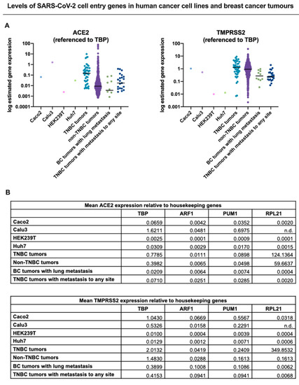
The COVID-19 pandemic has caused the need for prioritization strategies for breast cancer treatment, where patients with aggressive disease, such as triple-negative breast cancer (TNBC) are a high priority for clinical intervention. In this review, we summarize how COVID-19 has thus far impacted the management of TNBC and highlighted where more information is needed to hone shifting guidelines. Due to the immunocompromised state of most TNBC patients receiving treatment, TNBC management during the pandemic presents challenges beyond the constraints of overburdened healthcare systems. We conducted a literature search of treatment recommendations for both primary and targeted TNBC therapeutic strategies during the COVID-19 outbreak and noted changes to treatment timing and drugs of choice. Further, given that SARS-CoV-2 is a respiratory virus, which has systemic consequences, management of TNBC patients with metastatic versus localized disease has additional considerations during t he COVID-19 pandemic. Published dataset gene expression analysis of critical SARS-CoV-2 cell entry proteins in TNBCs suggests that the virus could in theory infect metastasized TNBC cells it contacts. This may have unforeseen consequences in terms of both the dynamics of the resulting acute viral infection and the progression of the chronic metastatic disease. Undoubtedly, the results thus far suggest that more research is required to attain a full understanding of the direct and indirect clinical impacts of COVID-19 on TNBC patients.

View on the web

Versatile Approach to Septonasal Deformity: Skyscraper Construction Based on an Old Relic

xloma.fota13 shared this article with you from Inoreader

SocialThumb.00001665.DC.jpeg

Introduction: Severely deformed noses usually harbor a combination of both bony pyramid and septal deformities. In this retrospective study, the authors aimed to evaluate our results of repair in patients with severe nasal deformities and importance of a versatile approach in these cases. Materials and Methods: A total of 32 cases with congenital or acquired (traumas or surgeries) severe nasal deformity were included in this retrospective study. Gender, age, etiology, reconstruction methods, complications, and results were recorded. Preoperative and postoperative pictures were compared; additionally, patients' reviews on the esthetic and functional outcomes were noted. Open approach, weak L-strut template preparation attached to a strong keystone skeleton and reconstruction with a stable L- or T-strut on this template were carried out in all cases. In addition, glabellar flaps were used in 2 cases to restore the contracted skin envelope and wide-angle L-shape cartilage grafts in 7 cases for extensive alar cartilage reconstruction. Results: Favorable esthetic and functional results were obtained in most of the patients. The postoperative problems were recorded as intranasal synechiae; costochondral graft displacement; residual external deviation; nostril asymmetry; residual alar, columellar and tip problems; and prolonged edema. Conclusions: Sufficient sizes and amounts of skin, mucosa, cartilage, and bone tissue must be available to plan versatile repair using flaps and grafts according to the needs of each patient. Preserved stability of the keylock area is substantial. The authors advocate construction of a new structure based on the native weakened skeleton free from the extrinsic and intrinsic forces is an effective method. EBM LEVEL 4 Address correspondence and reprint requests to Omer R. Ozerdem, MD, A-Plast Esthetic Plastic Surgery Center, Akasya Kent Kule A-1 K:24 D:124 34660 Acibadem, Istanbul, Turkey; E-mail: ozerdemor@yahoo.com Received 6 August, 2020 Accepted 4 December, 2020 The authors report no conflicts of interest. © 2021 by Mutaz B. Habal, MD.
View on the web