Αρχειοθήκη ιστολογίου

Αλέξανδρος Γ. Σφακιανάκης
ΩτοΡινοΛαρυγγολόγος
Αναπαύσεως 5
Άγιος Νικόλαος Κρήτη 72100
2841026182
6032607174

Δευτέρα 24 Οκτωβρίου 2022

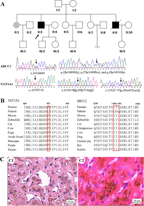
Concurrence of novel mutations causing Gilbert’s and Dubin–Johnson syndrome with poor clinical outcomes in a Han Chinese family

alexandrossfakianakis shared this article with you from Inoreader

10038_2022_1086_Fig1_HTML.png

Journal of Human Genetics, Published online: 24 October 2022; doi:10.1038/s10038-022-01086-1

Concurrence of novel mutations causing Gilbert's and Dubin–Johnson syndrome with poor clinical outcomes in a Han Chinese family
View on Web

Association between circulating 25‐hydroxyvitamin D metabolites and periodontitis: Results from the NHANES 2009‐2012 and Mendelian randomization study

alexandrossfakianakis shared this article with you from Inoreader

Abstract

Aim

This study sought to investigate associations of 25(OH)D metabolites with periodontitis, and to assess causality using Mendelian randomization (MR).

Methods

This study included 7,246 participants of the National Health and Nutrition Examination Survey, 2009–2012. The association of periodontitis with 25(OH)D metabolites was assessed using multivariable logistic regression analysis. 2-sample MR for 25(OH)D, 25(OH)D3, and C3-epi-25(OH)D3 with periodontitis (n = 17,353 cases/28,210 controls) was conducted. The principal analysis was inverse-variance-weighted approach. We controlled for horizontal pleiotropy using five additional methods.

Results

Based on the observational study, each 1-point increase in standard deviation of 25(OH)D lowered the risk of periodontitis by 15% (OR = 0.85, 95% CI: 0.78–0.93, P = 0.006) after multivariable adjustment. A similar relationship was observed between 25(OH)D3 and periodontitis (OR = 0.88, 95% CI: 0.80–0.97, P = 0.031). Furthermore, a potential nonlinear association was found between periodontitis with both 25(OH)D and 25(OH)D3. However, C3-epi-25(OH)D3 was not found to be associated with periodontitis risk. IVW-MR showed that periodontitis risk was not significantly associated with genetically increased levels of 25(OH)D (OR = 1.02, 95% CI: 0.90–1.16, P = 0.732), 25(OH)D3 (OR = 1.04, 95% CI: 0.93–1.17, P = 0.472), and C3-epi-25(OH)D3 (OR = 1.11, 95% CI: 0.87–1.41, P = 0.400). The pleiotropy-robust MR approaches yielded similar results after we had eliminated the variants with horizontal pleiotropy risk.

Conclusion

Cross-sectional observational analysis identified significant relationships between periodontitis with 25(OH)D metabolites, while findings based on MR study do not support a causal role.

This article is protected by copyright. All rights reserved.

View on Web

Κυριακή 23 Οκτωβρίου 2022

human monkeypox (hMPXV) re‐emergence: host immunity status and current vaccines landscape

alexandrossfakianakis shared this article with you from Inoreader

Abstract

Monkeypox virus is a member of the Orthopoxvirus genus and the Poxviridae family. Orthopoxviruses are among the most intricate animal viruses. The pathogenicity of human monkeypox infection has been emphasized in response to its recent emergence in non-endemic countries and the threat of bioterrorism. It is always necessary to take appropriate precautions in exposure to emerging or re-emerging infections. Here, we focus on the current state of the human monkeypox infection outbreak, research & development of immunity responses and clinical interventions to prevent and treat human monkeypox virus and other human poxviruses.

This article is protected by copyright. All rights reserved.

View on Web

Do cone‐beam computed tomography low‐dose protocols affect the evaluation of the temporomandibular joint?

alexandrossfakianakis shared this article with you from Inoreader

Abstract

Background

There is no established acquisition protocol based on scientific evidence for the acquisition of cone-beam computed tomography (CBCT) exams to evaluate the temporomandibular joint (TMJ).

Objectives

To evaluate the influence of acquisition protocols and jaw positioning on the diagnostic accuracy of TMJ condylar morphological alterations (CMA), dimension, position, and excursion.

Methods

Thirty-six TMJs on 18 dry skulls were imaged using a CBCT unit (OP300 Maxio, Instrumentarium, Tuusula, Finland) at two exposure settings (4.5 and 6.3 mA), three voxel resolutions (0.085, 0.125 and 0.280 mm), three jaw positions (concentric, anteriorized and posteriorized) and three jaw excursions (normoexcursion, hyperexcursion and hypoexcursion). The macroscopic anatomy examination and high-resolution CBCT images were used as ground truth for CMA. Twenty-five TMJs had at least one CMA with 11 healthy TMJs serving as controls. Three experienced oral and maxillofacial radiologists evaluated the parasagittal images for the presence of CMA, position, and excursion and measured dimensions. The area under the ROC curve, sensitivity, and specificity were calculated. Weighted Kappa (α=0.05) was used to determine intra- and inter-examiner reliability and comparisons between dependent variables analyzed by Analysis of Variance at an a prior level of significance of 0.05.

Results

The agreement of the evaluation of the position and excursion with the reference standard was high, independent of the protocol (range, 0.75-0.91). Various combinations of acquisition protocols and jaw position did not influence the CMA evaluation. Erosion was overdiagnosed in protocols with larger voxel sizes, and the detection of osteophytes greater in images with smaller voxel sizes. The anteroposterior dimension was greater in the open jaw position (p<0.05).

Conclusion

CBCT protocols using reduced radiation exposure from the CBCT machine evaluated in this study can be used to assess condylar morphology, dimension, position, and excursion, without compromising diagnostic performances for these parameters.

View on Web

Epigenetic upregulation of Schlafen11 renders WNT- and SHH- activated medulloblastomas sensitive to cisplatin

alexandrossfakianakis shared this article with you from Inoreader
Abstract
Background
Intensive chemotherapeutic regimens with craniospinal irradiation have greatly improved survival in medulloblastoma patients. However, survival markedly differs among molecular subgroups and their biomarkers are unknown. Through unbiased screening, we found Schlafen family member 11 (SLFN11), which is known to improve response to DNA damaging agents in various cancers, to be one of the top prognostic markers in medulloblastomas. Hence, we explored the expression and functions of SLFN11 in medulloblastoma.
Methods
SLFN11 expression for each subgroup was assessed by immunohistochemistry in 98 medulloblastoma patient samples and by analyzing transcriptomic databases. We genetically or epigenetically modulated SLFN11 expression in medulloblastoma cell lines and determined cytotoxic response to the DNA damaging agents cisplatin and topoisomerase I inhibitor SN-38 in vitro and in vivo.
Results
High SLFN11 expressing cases exhibited significantly longer survival than low expressing cases. SLFN11 was highly expressed in the WNT-activated subgroup and in a proportion of the SHH-activated subgroup. While WNT activation was not a direct cause of the high expression of SLFN11, a specific hypomethylation locus on the SLFN11 promoter was significantly correlated with high SLFN11 expression. Overexpression or deletion of SLFN11 made medulloblastoma cells sensitive and resistant to cisplatin and SN-38, respectively. Pharmacological upregulation of SLFN11 by the brain-penetrant histone deacetylase-inhibitor RG2833 markedly increased sensitivity to cisplatin and SN-38 in SLFN11-negative medulloblastoma cells. Intracranial xenograft studies also showed marked sensitivity to cisplatin by SLFN11-overexpression in medulloblastoma cells.
Conclusio ns
High SLFN11 expression is one factor which renders favorable outcomes in WNT-activated and a subset of SHH-activated medulloblastoma possibly through enhancing response to cisplatin.
View on Web

Σάββατο 22 Οκτωβρίου 2022

The added value of skeletal surveys in the initial evaluation of children diagnosed with Langerhans cell histiocytosis in the era of staging 18F‐FDG PET/CT: A retrospective study

alexandrossfakianakis shared this article with you from Inoreader

Abstract

Objective

Currently, there is no consensus protocol on the initial staging evaluation for Langerhans cell histiocytosis (LCH). Our institutional protocol consists of a skeletal survey and a whole-body positron emission tomography with 2-deoxy-2-[fluorine-18] fluoro-D-glucose integrated with computed tomography (FDG PET/CT) study. The utility of the PET/CT lies in its sensitivity in detecting osseous and extra-osseous lesions, and in determining the baseline metabolic activity of LCH lesions to assess treatment response. However, the added utility of the skeletal survey in staging LCH is unclear. Therefore, this study retrospectively assessed the added diagnostic value of skeletal surveys in patients with baseline PET/CTs for initial staging of LCH.

Methods

We retrospectively searched the medical records of all patients less than or equal to 18 years old at a large children's hospital (May 2013 to September 2021). The inclusion criteria were (a) biopsy-proven diagnosis of LCH and (b) initial staging PET/CT and skeletal survey performed less than or equal to 1 month apart. A blinded pediatric radiologist reviewed the skeletal surveys and another reviewed the PET/CTs in identifying LCH osseous lesions.

Results

Our study cohort consisted of 49 children with 86 LCH osseous lesions. In non-extremity locations, PET/CT identified 70/70 (100%) osseous lesions, while skeletal surveys detected 43/70 (61.4%) osseous lesions. In the extremities, PET/CT identified 13/16 (81.3%) osseous lesions, while skeletal surveys detected 15/16 (93.8%) osseous lesions.

Conclusion

Skeletal surveys increased the detection rate of osseous lesions in the extremities, but added no diagnostic value to the detection of osseous lesions in non-extremity locations. Therefore, we propose to abbreviate the skeletal survey to include only extremity radiographs.

View on Web

Histopathologically defined intestinal metaplasia in lesser curvature of corpus prior to Helicobacter pylori eradication is a risk factor for gastric cancer development

alexandrossfakianakis shared this article with you from Inoreader

Abstract

Background and Aim

Helicobacter pylori eradication has been shown to reduce the risk of gastric cancer (GC), with the number of eradication therapy cases on the rise. However, GC can still occur after successful treatment, and the histological differences prior to eradication in patients with and without GC are unclear. This study investigated the pre-treatment histological risk factors for GC development following eradication therapy.

Methods

We retrospectively enrolled consecutive adult patients diagnosed as having H. pylori infection between April 2004 and December 2018. Atrophy and intestinal metaplasia (IM) were histologically assessed according to the updated Sydney System. The operative link on gastritis assessment and the operative link on gastric intestinal metaplasia (OLGIM) were evaluated as well.

Results

Of the 247 patients analyzed in this study, 11 (4.5%) experienced GC after eradication therapy. Histological IM scores in the GC group were significantly higher at all gastric biopsy sites (p < .05), and the proportion of OLGIM III/IV stage was significantly greater in GC patients (81.8% vs. 31.8%, p < .01). For GC prediction, the area under the receiver operating characteristic curve for IM score at the lesser curvature of the corpus was the highest among all biopsy sites and not inferior to OLGIM results.

Conclusions

Patients with histological IM prior to H. pylori eradication, especially at the lesser curvature of the corpus, may be at elevated risk for GC development after eradication therapy and require close surveillance.

View on Web